Biomedical Engineering · Computational Neuroscience · Neural Imaging
Joel Jaramillo
B.E. Biomedical Engineering · The City College of New York · expected May 2028
Biomedical engineering undergraduate moving toward computational neuroscience and neural imaging.
Now
- Undergraduate Researcher, Bikson Lab (CCNY) Jul 2026 – present
- MRI and Machine Learning Research Assistant, ZVR Lab (University of Rochester) May 2026 – present
Surface
What you're looking at
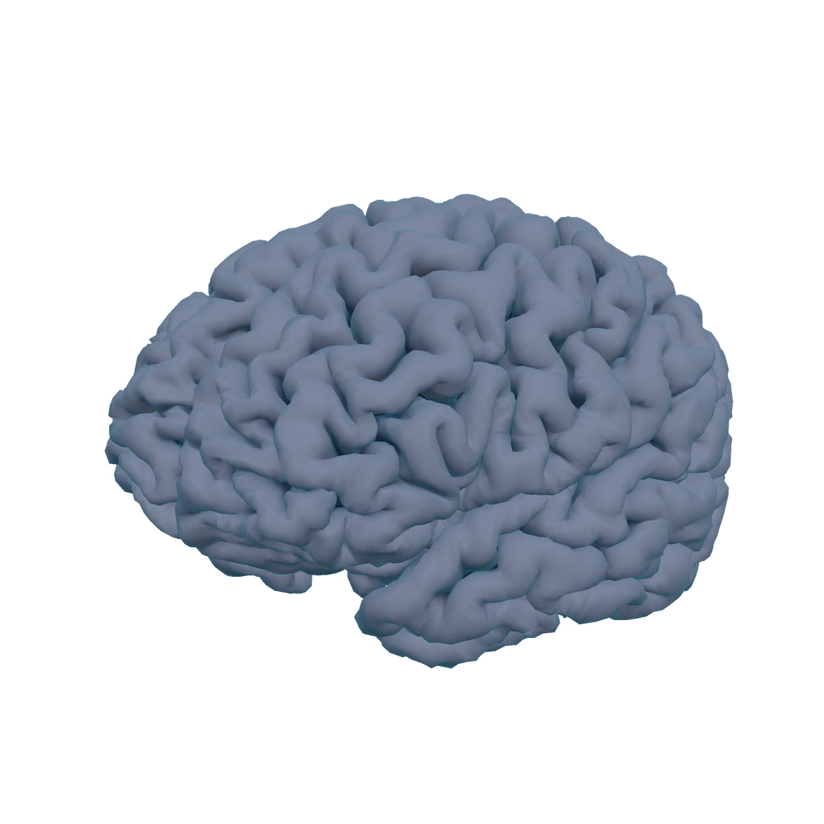
This is my brain's cortical surface, reconstructed from a research MRI with FreeSurfer 7.4.1. It's an anatomical visualization, not a medical image or a diagnosis.
Each page is a different operation on the same brain: slice through its deep structures, cut through it, read the atlas, or explore it freely. Switch the backdrop between the 3D model, my MRI slices, and a still image at the top right. Methods.
Featured case study
independentOne brain, twenty pipelines: how much do open-source neuroimaging tools agree?
I ran one T1-weighted MRI of my own brain through about 20 open-source segmentation tools and models, then compared what they report.
- neuroimaging
- structural-mri
- freesurfer
- segmentation
- reproducibility
- foundation-models