Atlas
About
B.E. Biomedical Engineering · The City College of New York · expected May 2028
I'm a biomedical engineering student at The City College of New York, moving toward computational neuroscience and neural imaging. My footing is engineering physics, circuits, and mechanics. I now apply it to MRI-based biomarker research and machine-learning analysis of brain data as a research assistant in the ZVR Lab at the University of Rochester, and to head modeling and electric-field simulation in the Bikson Lab at CCNY.
I'm comfortable across theory, Python data pipelines, and statistical inference, and I want to develop machine-learning and deep-learning methods for fMRI and brain-signal decoding. Outside research I train in combat martial arts.
Research interests
Areas I'm exploring, not areas of expertise.
- Neuroengineering Head modeling and electric-field simulation in the Bikson Lab.
- Neuromodulation Stimulation-electrode placement and field modeling.
- MRI imaging Structural MRI pipelines in the ZVR Lab, and my own scan.
- Machine learning and deep learning for brain data Classification in the ZVR Lab; EEG decoding in NeuroForge.
Skills
- Programming and data
- Python (NumPy, pandas, scikit-learn), MATLAB, Excel, Google Colab
- Neuroimaging and signals
- Structural MRI volumetrics, FreeSurfer, EEG, MNE-Python, MOABB, preprocessing and filtering
- Statistics
- ANCOVA, FDR and multiple-comparison correction, ROC/AUC, DeLong tests, effect sizes (Cohen's d, partial η²)
- ML and methods
- Classification (LDA, CSP), Riemannian geometry (pyRiemann), CNNs (EEGNet), cross-validation
- Engineering
- Circuit analysis, CAD (SolidWorks), COMSOL Multiphysics (FEA), Simpleware, oscilloscope and multimeter, fluids and material mechanics
Honors and affiliations
- NEUROCITY Summer Scholar, University of Rochester (2026)
- Dean's List, CCNY
- Member, Biomedical Engineering Society (BMES)
- In progress: Machine Learning Specialization (DeepLearning.AI)
Atlas of my cortex
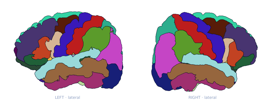
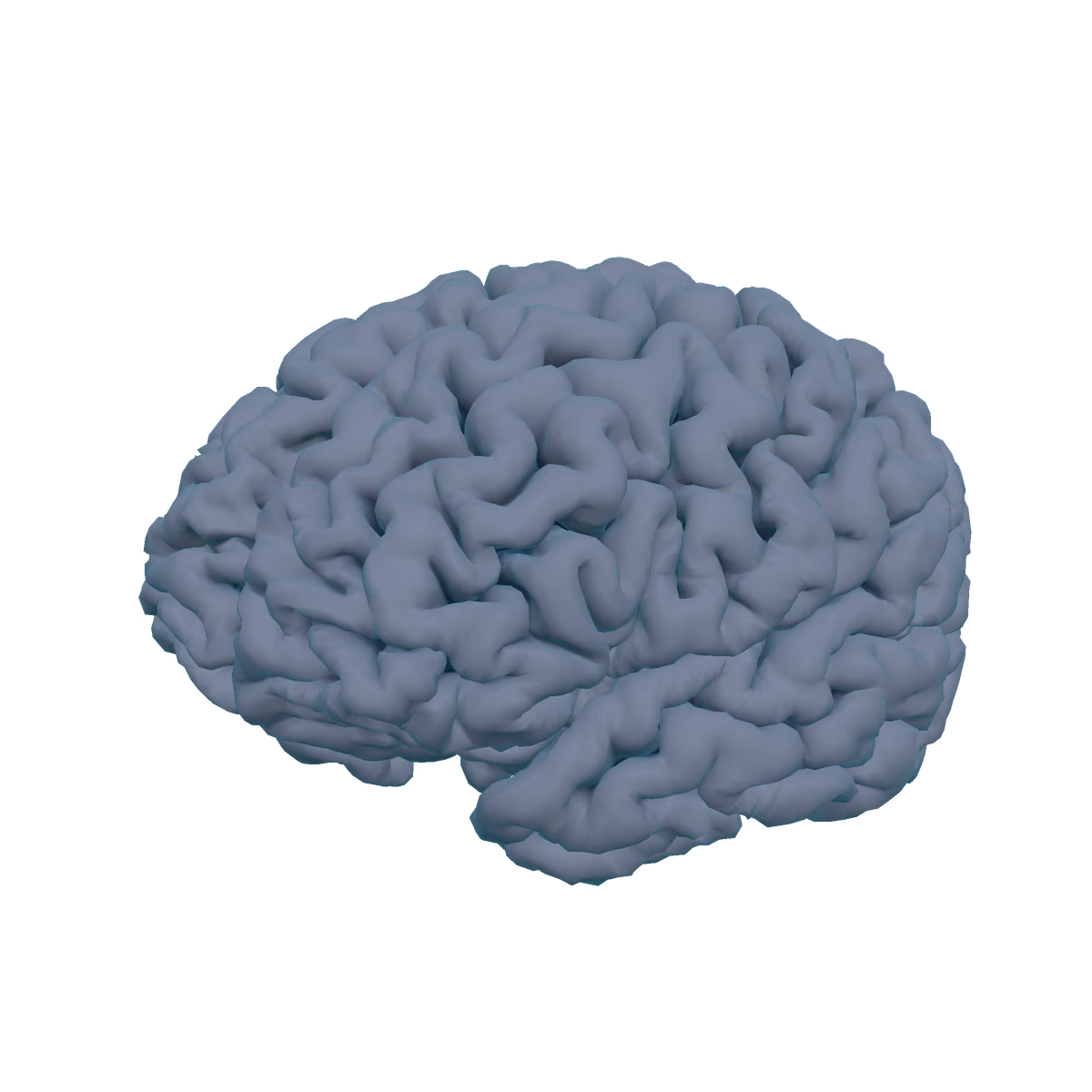
Desikan-Killiany (FreeSurfer aparc), 34 regions per hemisphere, colored with the FreeSurfer lookup table. Select a region on the brain or in the list. Descriptions use cautious "involved in" wording; brain regions work in networks, not alone. For the deep structures, open Explore.
No region selected. Choose one below, or click the brain.
Frontal (11)
- Caudal middle frontal
- Lateral orbitofrontal
- Medial orbitofrontal
- Paracentral lobule
- Pars opercularis
- Pars orbitalis
- Pars triangularis
- Precentral gyrus
- Rostral middle frontal
- Superior frontal
- Frontal pole
Parietal (5)
- Inferior parietal
- Postcentral gyrus
- Precuneus
- Superior parietal
- Supramarginal gyrus
Temporal (9)
- Banks of superior temporal sulcus
- Entorhinal cortex
- Fusiform gyrus
- Inferior temporal
- Middle temporal
- Parahippocampal gyrus
- Superior temporal
- Temporal pole
- Transverse temporal (Heschl's)
Occipital (4)
- Cuneus
- Lateral occipital
- Lingual gyrus
- Pericalcarine
Cingulate (4)
- Caudal anterior cingulate
- Isthmus cingulate
- Posterior cingulate
- Rostral anterior cingulate
Insula (1)
- Insula