Methods
How the brain on this site was made
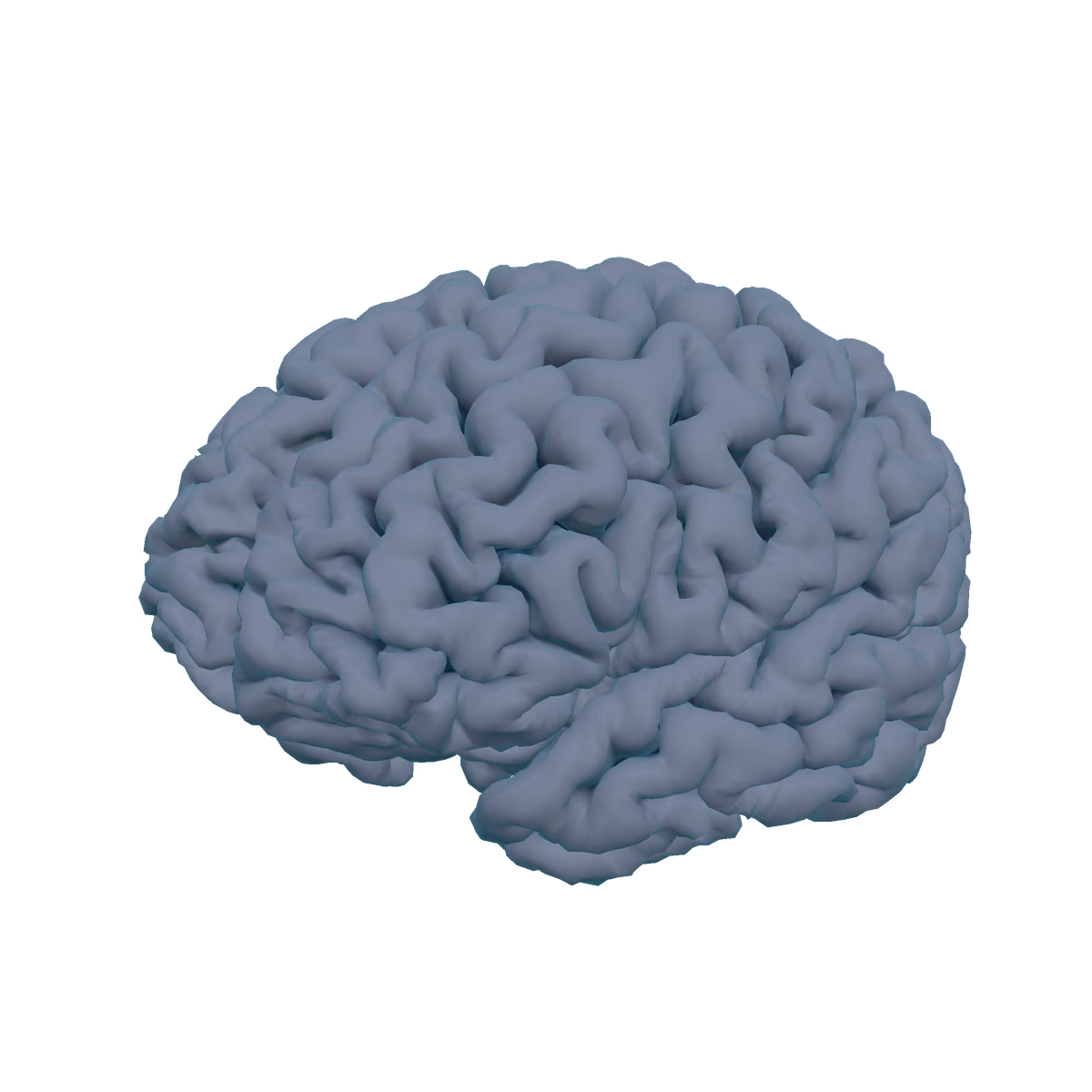
This is my brain's cortical surface, reconstructed from a research MRI with FreeSurfer 7.4.1. It's an anatomical visualization, not a medical image or a diagnosis.
Acquisition
- One 3D T1-weighted MRI, 1 mm isotropic, acquired on a Siemens scanner as part of a research acquisition (2026).
- This is a research scan, not a clinical one, and nothing on this site is a clinical reading of it.
Pipeline
- Reconstruction. FreeSurfer 7.4.1
recon-allproduced the pial (outer) and white (inner) cortical surfaces, the inflated surface, sulcal depth, curvature, cortical thickness, and the Desikan–Killiany parcellation. - Resampling.
mri_surf2surfresampled each hemisphere to an icosahedral mesh of order 6: 40,962 vertices and 81,920 triangles per hemisphere. All surfaces of a hemisphere share one vertex order, so the viewer can morph between them directly. - Web conversion. A build script (
scripts/build-brain.mjs) writes one glTF file per hemisphere. The pial surface is the base mesh; the inflated and white surfaces are morph targets. Positions are quantized to 14 bits and compressed with meshopt: 1.44 MB for both hemispheres, with a largest position error of 0.017 mm in a sampled round-trip check. - Rendering. three.js, through React Three Fiber, draws the surface with FreeSurfer-style shading from sulcal depth. Atlas colors come from the FreeSurfer lookup table (34 regions per hemisphere), desaturated by 20%.
- Deep structures.
scripts/build-deep.mjsturns FreeSurfer's 1 mm subcortical labels into meshes (surface nets, Taubin smoothing, meshopt simplification): Thalamus, Caudate nucleus, Putamen, Globus pallidus, Hippocampus, Amygdala, Nucleus accumbens, Ventral diencephalon, Lateral ventricle, Cerebellum, Brainstem. 21 meshes, 209 KB. Only shapes are shown, no volumes.
What each view shows
| Page | Operation | Data |
|---|---|---|
| Home | Surface | Pial surface, shaded by sulcal depth and curvature. |
| Research | Deep | Glass cortex with the subcortical structures inside, cut by a slice of the 1.5 mm T1 that moves as you scroll. Axial, coronal, or sagittal; the half on the + side of the slice (above, in front, or right) is cut away. |
| Projects | Section | An axial clipping plane. The cut face is filled from the surfaces themselves: the cortical ribbon lies between the pial and white surfaces, and the lighter fill is everything inside the white surface. No MRI intensities are shown. |
| About | Atlas | Desikan–Killiany regions. Selecting a region shows its FreeSurfer label, a plain-language description, and its mean thickness and surface area on my scan. |
| Explore | Viewer | Folded or inflated cortex (solid, glass, or hidden), deep structures you can select, and a slice plane that samples the 1.5 mm T1 as a 3D texture. The side of the brain facing you is clipped away at the plane. |
The coordinate readout is FreeSurfer tkr-RAS in millimetres, taken from the pial vertex nearest the pointer, even while the surface is inflated. The scale bar is measured at the center of rotation.
What this site does not show
- No MRI slices or volumes, no face, and no DICOM metadata. Only cortical surfaces are published.
- No health information: no normative comparisons, z-scores, percentiles, brain-age estimates, screening outputs, or incidental findings.
- Region descriptions are general neuroscience, not statements about my brain. Regions work in networks, and "involved in" is deliberate.
- Thickness and area values are FreeSurfer's anatomical measurements, shown without comparison to anyone else.
Privacy choices
- The scan date is shown as a year only.
- Raw and full-resolution imaging data never enter the website's repository. The site ships only resampled surfaces.
- The site uses no cookies. Your backdrop and light-mode choices are stored only in your own browser.
Background image
The faint background is a fluorescence micrograph of rat cortical neurons expressing GFP, by ManuelSchottdorf, used under CC BY-SA 4.0 (cropped, converted to grayscale, and recolored; this adaptation is also CC BY-SA 4.0). Source. It is someone else's image, shown for atmosphere at 16% opacity, and it is not my data.

Performance tiers
- Full 3D on desktop browsers with WebGL2.
- Light 3D on phones, low-memory devices, and Save-Data connections: the 3D model and MRI slices load only when you ask, there's no idle rotation, and pre-rendered slices replace the live cut.
- No 3D without WebGL2 or when you choose the Still backdrop: a still image and a 2D atlas. MRI slices still work, because they're drawn in 2D. Every page stays fully readable.